
Understanding Actinic Keratosis Treatment: A Comprehensive Guide
A guide to understanding, treating, and preventing Actinic Keratosis using proven medical and natural approaches.
Actinic keratosis, a common precancerous skin condition, affects millions of people worldwide. These scaly patches typically appear on sun-exposed areas of the body and can potentially develop into skin cancer if left untreated. Understanding actinic keratosis treatment options is crucial for managing this condition effectively and preventing its progression to more serious forms of skin cancer, including melanoma.
What is Actinic Keratosis?
Actinic keratosis, also known as solar keratosis, is a skin condition that develops on sun-exposed areas of the body due to prolonged exposure to ultraviolet (UV) radiation. Actinic keratoses typically appear on the face, ears, scalp, neck, forearms, and hands of individuals with a history of cumulative sun exposure.
Definition and Causes
Actinic keratosis is characterised by the presence of rough, dry, or scaly patches on the skin that may be skin-coloured, pink, red, or brown. These lesions are caused by the cumulative effects of UV radiation on the skin, which triggers pathological changes in the epidermal keratinocytes by disrupting regulatory pathways involved in cell growth and differentiation. This disruption leads to inflammation, immunosuppression, and the intraepidermal proliferation of dysplastic keratinocytes, which are the precursors of actinic keratosis.
Risk Factors
Several factors can increase an individual’s risk of developing actinic keratosis. Age is a significant risk factor, as actinic keratoses are more common in people over 40 due to the high cumulative lifetime exposure to the sun and inadequate sun protection measures. Fair-skinned individuals with light hair and eyes are more susceptible to the damaging effects of UV radiation and have a higher risk of developing actinic keratosis. Additionally, people living in geographic locations closer to the equator, those with a history of sunburns, and individuals with weakened immune systems due to medical conditions or immunosuppressive medications are at an increased risk.
Signs and Symptoms
Actinic keratoses can present in various forms, such as scaly, erythematous macules, papules, plaques, or cutaneous horns. The lesions may be skin-coloured, red, pink, grey, yellow, brown, or tan, and their surface can be rough, dry, and scaly. In some cases, actinic keratoses may cause symptoms such as itching, burning, tenderness, or bleeding when traumatised. The surrounding skin may also exhibit signs of chronic sun damage, such as wrinkling, mottled pigmentation, or telangiectasias.
It is crucial to recognize the signs and symptoms of actinic keratosis and seek prompt medical attention, as these lesions have the potential to progress into skin cancers. Early detection and treatment of actinic keratosis can help prevent the development of skin cancer and minimise the risk of complications. Individuals with a history of sun exposure should undergo regular skin examinations by a dermatologist to identify and manage actinic keratoses effectively.
Diagnosis and Early Detection
Early detection and accurate diagnosis of actinic keratosis are crucial for effective treatment and prevention of potential progression to skin cancer. Dermatologists play a vital role in identifying these precancerous lesions through clinical examination and diagnostic techniques.
Clinical Examination
A thorough clinical examination is the first step in diagnosing actinic keratosis. Dermatologists carefully inspect the skin, particularly in sun-exposed areas such as the face, scalp, ears, neck, forearms, and hands. They look for characteristic features of actinic keratosis, including rough, scaly patches or lesions that may be skin-coloured, pink, red, or brown. The lesions are usually small, ranging from a few millimetres to a centimetre in diameter. Dermatologists also assess the surrounding skin for signs of sun damage, such as wrinkling, mottled pigmentation, or telangiectasias.
During the examination, dermatologists may palpate the lesions to determine their texture and thickness. Actinic keratoses often have a sandpaper-like feel due to the presence of hyperkeratosis. The number, size, and distribution of lesions are also noted, as multiple actinic keratoses may be present within a single anatomic area.
Dermoscopy
Dermoscopy is a non-invasive diagnostic tool that allows dermatologists to examine skin lesions in greater detail. It involves the use of a handheld device called a dermatoscope, which magnifies the skin and provides illumination to visualise subsurface structures. Dermoscopy can aid in the differentiation of actinic keratosis from other benign or malignant skin lesions.
Dermoscopic features of actinic keratosis may include a “strawberry pattern” characterised by an erythematous background with prominent follicular openings surrounded by a white halo. Other dermoscopic findings may include surface scales, fine linear-wavy vessels, and shiny white streaks. Pigmented actinic keratoses may exhibit additional features such as brown or gray dots, globules, or a pigmented network.
Dermoscopy has been shown to improve the diagnostic accuracy of actinic keratosis, particularly when used by experienced dermatologists. It can help identify early or subtle lesions that may be difficult to appreciate with the naked eye.
Skin Biopsy
In certain cases, a skin biopsy may be necessary to confirm the diagnosis of actinic keratosis or rule out the presence of a skin cancer. A biopsy involves removing a small sample of the suspicious lesion for histopathological examination under a microscope.
There are different types of skin biopsies, including shave biopsy, punch biopsy, and excisional biopsy. The choice of biopsy technique depends on the size, location, and clinical suspicion of the lesion. Shave biopsies are commonly performed for superficial lesions, while punch or excisional biopsies may be indicated for thicker or more concerning lesions.
The biopsy sample is processed and examined by a dermatopathologist who evaluates the microscopic features of the tissue. Histologically, actinic keratosis is characterised by the presence of atypical keratinocytes in the lower layers of the epidermis, along with hyperkeratosis and parakeratosis in the stratum corneum.
Early detection of actinic keratosis through regular skin examinations and prompt diagnosis is essential for timely intervention and prevention of potential malignant transformation. Dermatologists play a crucial role in educating patients about the importance of sun protection, self-skin examinations, and the need for regular follow-up visits to monitor for the development of new or changing lesions.
By combining clinical examination, dermoscopy, and skin biopsy when necessary, dermatologists can accurately diagnose actinic keratosis and develop individualised treatment plans to address these precancerous lesions effectively. Early intervention can help prevent the progression of actinic keratosis to invasive squamous cell carcinoma and minimise the risk of skin cancer.
Treatment Options for Actinic Keratosis
Several treatment options are available for actinic keratosis, including topical medications, cryotherapy, photodynamic therapy, and surgical procedures. The choice of treatment depends on various factors such as the number, size, and location of the lesions, as well as patient preferences and overall health status.
Cryotherapy
Cryotherapy, also known as cryosurgery, is a commonly used procedure for treating individual actinic keratosis lesions. It involves the application of liquid nitrogen to the affected area, causing the formation of ice crystals within the cells, leading to their destruction. The procedure is performed in the office setting and does not require anaesthesia.
The effectiveness of cryotherapy depends on factors such as the duration of freezing, the number of freeze-thaw cycles, and the size and thickness of the lesion. Thicker, hypertrophic lesions may require longer freezing times or multiple treatment sessions.
Common side effects of cryotherapy include pain, redness, swelling, and blistering at the treatment site. These usually resolve within a few days to weeks. Rarely, cryotherapy may cause scarring or permanent changes in skin pigmentation, especially in individuals with darker skin tones.
Cryotherapy is an effective treatment option for isolated actinic keratosis lesions, with reported clearance rates ranging from 39% to 83%, depending on the treatment protocol. However, it may not be suitable for treating large areas with multiple lesions or field cancerization, as it can be time-consuming and may result in a less favourable cosmetic outcome compared to field-directed treatments.
Topical Medications
Topical treatments are often the first-line approach for non-hyperkeratotic, non-hypertrophic, visible or palpable actinic keratoses (grade I, II). These medications are applied directly to the affected sun-exposed areas and work by selectively destroying the abnormal cells while minimising damage to the surrounding healthy skin.
Commonly used topical agents include imiquimod, 5-fluorouracil (5-FU) and tirbanibulin. Imiquimod is an immune response modifier that stimulates the body’s immune system to fight the precancerous cells. It is available in different concentrations (5%, 3.75%, and 2.5%) and is typically applied two to three times per week for several weeks. 5-FU is an antimetabolite that interferes with DNA synthesis, leading to cell death. It is applied once or twice daily for two to four weeks.Tirbanibulin, a recently approved topical treatment, inhibits tubulin polymerization and protein kinase signalling, leading to the death of rapidly dividing cells. It is applied once daily for five consecutive days.
Topical aldara – a cream applied to broad area of actinic keratosis in a slow process over many weeks. This cream stimulates your immune system to attack and remove the actinic keratoses.
These topical treatments can cause local skin reactions such as redness, swelling, itching, and crusting, which usually resolve within a few weeks after completing the treatment course. Patients should be educated about the expected side effects and the importance of adhering to the prescribed treatment regimen to achieve optimal results.
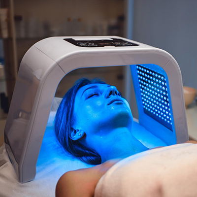
Photodynamic Therapy
Photodynamic therapy (PDT) is a minimally invasive procedure that combines the use of a photosensitizing agent with light exposure to selectively destroy precancerous cells.
It’s a clever combination of a special cream and a targeted light source that work together to wipe out precancerous cells while leaving healthy skin unharmed.
Here’s how it works:
- Apply the “tracker”: First, a cream containing a photosensitizing agent (think of it as a tracker that sticks to the bad cells) is applied to the affected area.
- Let it soak in: The cream is left on for a while to allow the “tracker” to be absorbed by those abnormal cells.
- Activate with light: Next, the area is exposed to a specific type of light, which activates the “tracker” and triggers a reaction that destroys the targeted cells.
PDT can tackle individual spots or larger areas of sun damage.
Daylight PDT: A Brighter Approach
There’s even a newer version called daylight PDT that uses natural sunlight instead of artificial light. This means a shorter waiting time and less discomfort during the treatment.
While PDT is generally safe, you might experience some temporary redness, swelling, or a burning sensation afterward. But these side effects usually fade quickly.
PDT offers a minimally invasive and highly effective way to combat actinic keratosis, giving you a brighter outlook on your skin’s health.
Surgical Options for Actinic Keratosis
When dealing with stubborn or suspicious actinic keratosis (AK) spots, sometimes you need a more direct approach. That’s where surgical procedures come in. These techniques involve removing the troublesome lesions with precision, giving you immediate results and peace of mind.
Think of it like this:
- Curettage: This is like carefully scraping away the unwanted spot with a specialised tool, often combined with a little electrical zap to stop any bleeding and ensure no abnormal cells are left behind.
- Shave Excision: Imagine a dermatologist skillfully shaving off the lesion with a scalpel, leaving a smooth surface.
- Surgical Excision: This is a bit more involved, where the entire lesion and a small safety margin of healthy skin are removed. It’s usually reserved for spots that look suspicious or haven’t responded to other treatments.
The upside? These procedures get rid of the lesion right away, and your doctor can even examine the removed tissue to make sure everything is okay. The downside? There’s a chance of scarring, and it might not be the best option for lots of spots or larger areas.
Stopping AK Before it Starts: Prevention is Key
The best way to deal with actinic keratosis? Prevent it in the first place! Since the sun’s harmful UV rays are the main culprit, sun protection is your superpower:
- Dress for the occasion: Cover up with protective clothing, a wide-brimmed hat, and sunglasses.
- Slather on the sunscreen: Make sunscreen your daily BFF. Choose a broad-spectrum sunscreen with an SPF of 30 or higher and reapply it regularly, especially after swimming or sweating.
- Seek the shade: When the sun’s at its strongest (usually between 10 am and 4 pm), find some shade to hang out in.
Regular skin checks are like your early warning system. Your dermatologist can spot suspicious spots early on, and you can also do self-exams at home to keep an eye on your skin. Remember, catching AK early can prevent it from turning into something more serious, like skin cancer.
Conclusion
To wrap up, understanding and managing actinic keratosis is crucial to prevent its progression into skin cancer. This comprehensive guide has explored various aspects of this condition, from early detection to long-term management, covering different treatment approaches for various types of actinic keratosis. The information provided aims to help readers make informed decisions about their skin health and take proactive steps to protect themselves.
Ultimately, preventing actinic keratosis through sun protection and regular skin checks is key to reducing the risk of skin cancer. By adopting these preventive measures and staying vigilant, individuals can minimise the chances of developing actinic keratosis and its potential consequences. Remember, early detection and prompt treatment are essential to maintain healthy skin and prevent more serious complications down the road.
FAQs
What are some effective preventative measures for actinic keratosis?
To prevent actinic keratosis, adopt these key habits:
- Apply sunscreen daily and reapply it every two hours.
- Avoid tanning.
- Wear protective clothing such as wide-brimmed hats, sunglasses, long sleeves, and pants.
- Seek shade when outdoors.
- Regularly perform self-examinations of your skin.
What are the common treatment options available for actinic keratosis?
Actinic keratosis can typically be treated in one or two office visits. Common treatments include:
- Cryosurgery: This involves freezing the AK lesions to remove them.
How many cryotherapy sessions are usually required to treat actinic keratosis?
For actinic keratosis, treatment generally involves one session of cryotherapy, which includes a freeze–thaw cycle to effectively treat the sun-damaged lesions.